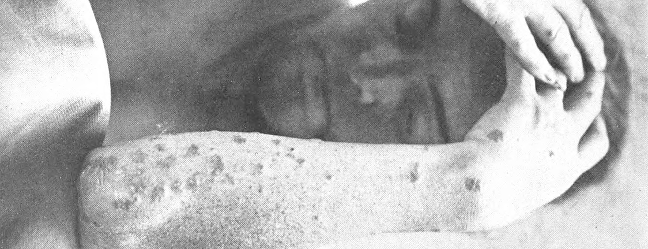
Dermographic Opacities: First Response

Sean Purcell’s "Dermographic Opacities" takes us into and through (even behind) a series of late nineteenth- and early twentieth-century clinical dermatological photographs whose subjects – or rather, whose subjected and objectified bodies – represent the production of medical knowledge via the exploitation of marginalized people. The result is an emphatic and at times uncomfortably necessary reckoning with the power imbalances that have marked medical "progress" over the past century and more. "What is to be done," Purcell asks, "with medical knowledge that relies on an exploited subject?" Well-known, yet still under-appreciated, cases of medical exploitation including Saartjie ‘Sarah’ Bartmann, Henrietta Lacks, the Tuskegee experiment, and the abuse of concentration camp inmates for Nazi experimentation make a quick appearance here, but Purcell’s real focus is on the anonymous – yet not anonymized – bodies and skins that appear in medical textbook photographs to illustrate a variety of dermatological conditions. The anonymous nature of the people in the photographs reduces their humanity, presenting them for inspection as non-person exhibits or mere objects whose purpose is to serve what Purcell calls the "production of knowledge claims." At the same time, because no attempt was made to conceal or anonymize the personal identities of the photographed persons from those who would later gaze with instinctual distaste and/or clinical curiosity upon their spotted, deformed, ulcerous, and pustulated bodies and faces, the "images fortify assumptions concerning race, gender, sexuality, ability, and class". The people in the photographs thus are turned into "others" who neither matter nor exist beyond what their maladies might teach us.
Purcell’s choice of dermatological photographs is an effective and interesting one. As he notes, he chose dermatology "because of its focus on the skin, a psychosomatic register that can signify difference as well as emotional and physical feeling." Meanwhile, his focus on photographs, which he effectively alters to highlight both the coercion exercised over the subjects and the physiognomy of their diseases, makes clear the "ways of thinking [that] maintain forms of othering, particularly concerning race, ability, and gender" and forces us to reconsider "the epistemic assumptions concerning what is known, what should be known, and how it can be known."
Other categories of medical photographs respond to similar arguments, both buttressing Purcell’s larger purpose and offering additional lines of enquiry. Jean-Martin Charcot’s photographs of demonstratively "hysterical" women at the Pitié-Salpêtrière Hospital in Paris – some of which were later collected and published in the multivolume Iconographie photographique de la Salpêtrière: service de M. Charcot (Paris: Progrès médical, 1877–80) – served not only to build and enhance Charcot’s own reputation as a visionary and educator in the emerging fields of neurology and psychology, but also to cement (despite numerous debates) the acceptance of hysteria as a legitimate medical diagnosis and thus a tool to further marginalize those who did not conform to social expectations. The women in Charcot’s many photographs are also objectified representations of a newly (re)defined clinical medical condition, most of them poor, anonymous, and shown at their most vulnerable with little attempt to conceal their identities. Other similarly problematic examples include photographs of female patients diagnosed with anorexia nervosa and published in medical journals, of "racial patholog[ies]" published in Rudolph Mata’s "Surgical Peculiarities of the American Negro" (1896), and of psychiatric patients inserted into case books, textbooks, and academic journals.[1] In these cases and many more, medical photography signalled scientific progress in two ways – as a technology and as an objective, realistic representation of clinical diagnostic practices – while simultaneously, for the subject/object of the photograph, contributing to what Erin O’Connor calls "the erasure of personhood."[2]There is also an extremely hazy, perhaps purposefully blurred, distinction between the medical and clinical value of photographs depicting people described as "medical oddities" – non-normative bodies in modern parlance – and the voyeuristic exploitation of these same people when showcased as exhibits in "freak shows."[3]
Two Sets of Observations
I’d like to return to Purcell’s focus on the skin and offer two very different sets of observations as a contribution to thinking about biases in images of disease. The first relates to diagnostics and race and the second to the misuse of much older (i.e. pre-photography) images to portray diseases of the past.
There is a perhaps ironic twist to the racial assumptions that often underlay medical photography of dermatological conditions. Certain conditions that embody moralistic undertones, especially those related to sexually transmitted or particularly disfiguring diseases, are often portrayed in historical medical photographs and films as afflictions of the racialized: the syphilitic Black man and the native Hawai’ian "lepers" shown in Purcell’s visual essay are but two examples among many. Yet as our recent (and ongoing) experience with COVID-19 reminds us somewhat starkly, diseases often produce different symptoms on white and coloured bodies. Clinical knowledge, practice, and diagnosis in the West, or the Global North, is typically – and long has been – based on how symptoms appear on white bodies, leaving physicians and other health practitioners "better equipped to recognise and diagnose conditions as they appear on a white body than they are on a non-white body."[4] Everything from lesions to rashes and pale or otherwise discoloured skin is described in medical textbooks as they appear on white skin. This in turn leads to a significant bias in terms of practitioners’ ability to diagnosis the same conditions on non-white skin, and to disproportionately higher and more severe infection (and ultimately mortality) rates among black and brown bodies. The lack of racial and ethnic diversity in medical research and clinical trials that would help to overturn these biases is well known,[5] as are the longstanding, racially based socio-economic inequalities that permeate public health and access to healthcare to this day. And yet, non-white bodies have been more likely to be exploited, either as embodied subjects/objects of medical education and research or in medical photography designed to depict the worst cases of dermatological conditions – essentially those left untreated for so long that they became the showcased epitome of how this or that skin disease could, ultimately, present itself.
If one searches for modern clinical photographs of bubonic plague online, what typically appear are racialized bodies. Granted, except for the western United States, the disease is now largely absent from the West/Global North, and is relatively easily treated with antibiotics. In the past, of course, this was not the case. Through three multi-century and overlapping pandemics, much of Eurasia, Sub-Saharan Africa, and later the Americas, faced repeated plague epidemics. Clinical medical photography of the Third Pandemic (began c.1855) emerges largely from India, Mongolia, Brazil, and, more recently, Madagascar.[6] For the pre-photograph era, though, we are forced to look to illustrations to tell us how the dermatological manifestations of plague might have appeared. And herein lies a huge problem. Thanks to multiple digitisation projects – and the reposting of digitised images online – scholars and the general public now have access to a greater variety and number of historical images of plague than they ever have before. However, seemingly innocuous actions, such as inaccurate modern captioning, have actually changed the underlying meaning of numerous medieval and early modern images into something that they were never meant to represent. Furthermore, the context in which these images first appeared make it clear that they come from non-medical sources (i.e., chronicles or religious works) that had no ostensible pedagogical or clinical medical function; there is, as a result, no explicit reason to label the disease being portrayed in medical terms. In short, the images are not medical illustrations that can be equated to medical photographs, but instead were intended as visual contributions to a narrative about something else entirely – the celebration of a religious life, for example, or instructions for leading one.
I discuss this problem in more detail elsewhere,[7] but want to emphasize here that almost all of the easily obtained online images of medieval and early modern plague symptoms on people’s skin do not, in fact, depict plague. What we see, instead, are cropped, decontextualized, and mislabelled images of either some other disease (most often leprosy) or biblical analogies of the 10 Plagues of Egypt (of which plague, the disease, was noticeably absent). This matters because in continuously re-using these images incorrectly, we are perpetuating a false narrative about people’s experience with disease in the past. We are also seriously misrepresenting the physiognomy of disease for diagnosis and treatment – what plague looked like on the skin of those who suffered from it then and, by extension, of those who continue to suffer from it today. While such illustrations do not present the same kind of subjected objectification that we encounter with the exploited and marginalized bodies of medical photography, we do nevertheless face the problem of misguided "knowledge" production and dissemination. No single individual is anonymized, erased, and rendered non-human by misreading historical, pre-photographic disease images; yet in doing so millions of people’s experiences with that disease and its symptoms are reduced to the level of a caricature that significantly "others" those who still encounter plague today.
This brings us back to Purcell’s "Dermographic Opacities" and the problem that arises when the exploitative photographs are made opaque. As Purcell notes,
"When I isolate the disease to the degree where the disease is wholly and totally itself (in the ideal of the objective sciences), its meaning becomes increasingly, impossibly abstract... If I try to isolate the disease, I find myself over abstracting the object. Without its ground—the body—the disease loses its figural power."
In short, what Purcell achieves in the end by removing the entire image except for the disease itself is relevant – because it achieves nothing that helps to help us to understand dermatological diseases.
Perhaps the answer, then, is not to attempt to make the dermatological photographs opaque by removing everything except the disease symptoms, but rather, as Katherine D.B. Rawling has done with psychiatric photographs, to find a way to restore agency and selfhood to the people in them.[8] Their appearance in textbooks and articles is distressing because they were not cropped to disguise personal identities, as they would be today. De-humanising as such photos are now considered, though, in their time they may have been considered revolutionary for their pedagogical value. In that respect, the photos themselves are contributive stepping-stones to knowledge making, however distasteful and marginalising they are to us. They do have something to teach us, much more perhaps than simply as photographs of people with disfiguring skin ailments.
Taking this alternative approach, we can begin to re-recognise the subjects of medical photographs as individual persons. Their identities may be lost to us historically, but resituating them in their original context and recognizing them as individuals who lived lives marked not only by marginalization, exploitation, and disease, but also by everything else that makes us human could go some way to mitigating the visceral discomfort we experience in viewing their subjected bodies today.
Cover Image Lennep, Henry J. van, "Bible Lands, their modern customs and manners illustrative of Scripture" p218 British Library
Masthead Image Darier, J., 1920. A Text-Book of Dermatology. Lea & Febiger, Philadelphia & New York. p.50.
Erin O’Connor, "Pictures of Health: Medical Photography and the Emergence of Anorexia Nervosa,"Journal of the History of Sexuality 5, no. 4 (1995): 535–72; Stephen C. Kenny, "Capturing Racial Pathology: American Medical Photography in the Era of Jim Crow," American Journal of Public Health 110, no. 1 (2020): 75–83; Katherine D. B. Rawling, "‘She sits all day in the attitude depicted in the photo’: Photography and the Psychiatric Patient in the Late Nineteenth Century," Medical Humanities 43, no. 2 (2017): 99–110. ↩︎
Erin O’Connor, "Camera Medica: Towards a Morbid History of Photography," History of Photography 23, no. 3 (1999): 232–44 (p.235). ↩︎
There is a large literature here; see, among many others, Robert Bogdan, Freak Show: Presenting Human Oddities for Amusement and Profit (University of Chicago Press, 1990) and Picturing Disability: Beggar, Freak, Citizen and Other Photographic Rhetoric (Syracuse University Press, 2012); Anna Kerchy and Andrea Zittlau, eds., Exploring the Cultural History of Continental European Freak Shows and ‘Enfreakment’ (Cambridge Scholars Publishing, 2013); Nadja Durbach, "‘Skinless Wonders’: Body Worlds and the Victorian Freak Show," Journal of the History of Medicine and Allied Sciences 69, no. 1 (2014): 38–67. ↩︎
For a longer discussion of this point, and of the relationship between colours and diseases, see Nükhet Varlık, "Colours of Disease and Death in the Early Modern Ottoman Cultural Imagination," in Death and Disease in the Medieval and Early Modern World: Perspectives from Across the Mediterranean and Beyond, edited by Lori Jones and Nükhet Varlık (York Medieval Press, 2022). ↩︎
R. C. Rabin, ‘Dermatology Has a Problem With Skin Color’, The New York Times (30 August 2020) (last accessed 12 May 2022). ↩︎
See Christos Lynteris, ed., Plague Image and Imagination from Medieval to Modern Times (Palgrave Macmillan, 2021) and "Visual Representations of the Third Plague Pandemic," an ERC-funded project based at CRASSH, University of Cambridge, and The University of St Andrews, running from 2013 until 2018. ↩︎
Lori Jones and Richard Nevell, "Plagued by Doubt and Viral Misinformation: The Need for Evidence-based Use of Historical Disease Images," The Lancet Infectious Diseases 16, no. 10 (Oct. 2016): 235–40. ↩︎
Rawling, "‘She sits all day.’" ↩︎
